The use of x-rays in dentistry is considered a necessary component of a thorough dental examination. Problems cannot be treated without first being detected. Diagnosing and treating dental problems at an early stage can prevent pain, promote future dental health, and save time and money. In addition to cavities, x-rays allow us to detect an abscess (infection), cysts, tumors, extra and/or missing teeth, and other clues to overall dental development.
In pediatric dentistry, we sometimes get the comment “they’re just baby teeth”. Parents are often surprised when we inform them their child may be teething into their teenage years. Parents can appreciate and understand the need for radiographs when we take the time to educate them that the health of primary (baby) teeth can affect future dental health and prevent pain. Did you know that dental problems account for nearly 2 million missed school days each year? Even if a child with tooth pain is able to make it to school, you can imagine that pain would interfere with concentration and learning. We like to use x-ray technology to identify cavities when they are small and limited to the outer layers of the tooth, before the cavity has a chance to cause any pain.
Generally, children require radiographs more often than adults since the enamel of baby teeth is much thinner than permanent teeth and their mouths grow and change rapidly. We at Sweet Tooth Pediatric Dentistry understand concerns parents have with regard to exposing children to x-rays. Therefore, we will only take x-rays that are necessary to provide our patients with optimal care that we would also recommend for our own children and families. We follow the American Academy of Pediatric Dentistry’s recommendation for the type and frequency of x-rays and parents are always informed before any radiographs are taken. Our state-of-the-art digital x-ray equipment allows us to obtain the best diagnostic images using the smallest amount of radiation possible. The combination of this digital equipment along with a lead body apron and thyroid collar shield assures a minimal amount of radiation exposure.
Below is a summary of the types of x-rays we taken in our office.
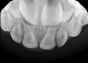
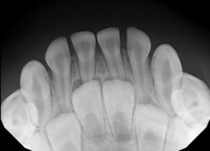
OCCLUSAL X-RAYS: Images of the top front and/or bottom front primary teeth to identify decay, infection, trauma, extra and/or missing teeth.
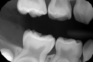
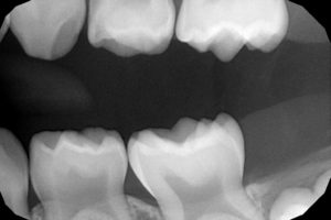
BITEWING X-RAYS: Images of the back teeth most widely used to detect decay in between teeth. These x-rays are needed when primary molars first begin to touch each other.
PERIAPICAL X-RAY: Image of a tooth, its root, and surrounding bone commonly used to diagnose potential problems associated with the root of a tooth such as abscesses and fractures.
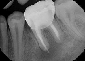
PANORAMIC X-RAY: Image of the entire mouth including all teeth (including those that are not erupted yet), the upper and lower jaws, temporomandibular joints (TMJ), nasal area, and sinuses. Taken at specific phases to monitor growth and development, identify extra and missing teeth, impacted teeth, as well as cysts and tumors.

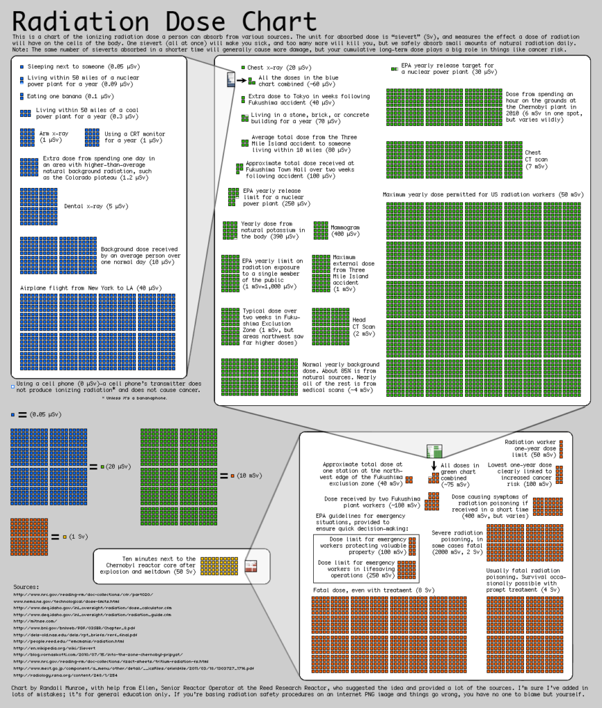
As you are aware, we are exposed to natural background radiation from the earth, sun, moon, and stars. You may find this radiation comparison chart interesting as it relates various other sources of radiation and the dental x-ray. For instance, we get more radiation on an airplane flight from New York to LA than from a dental x-ray!
We at Sweet Tooth Pediatric Dentistry welcome any questions you may have related to your child’s x-rays. Patient safety is our top priority and we pride ourselves on offering a conservation radiation schedule that is both helpful and safe.
Amie Beaulieu
Pediatric Dental Hygienist
Sweet Tooth Pediatric Dentistry
583 Saybrook Road
Middletown, CT 06457
(860) 347-4681
www.sweettoothkids.com
